Flow cytometric analysis of Amyloid beta precursor like protein 2 expression in HepG2 cells using anti-Amyloid beta precursor like protein 2 antibody (Cat#3919, 1:2,000). Green, isotype control; red, Amyloid beta precursor like protein 2.
Immunocytochemical staining of HepG2 cells with anti-Amyloid beta precursor like protein 2 antibody (Cat#3919, 1:1,000) . Nuclei were stained blue with DAPI; Amyloid beta precursor like protein 2 was stained magenta with Alexa Fluor® 647. Images were taken using Leica stellaris 5. Protein abundance based on laser Intensity and smart gain: High. Scale bar, 20 μm.
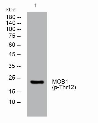
Western blotting analysis using anti-amyloid beta precursor like protein 2 antibody (Cat#3919). Total cell lysates (30 μg) from various cell lines were loaded and separated by SDS-PAGE. The blot was incubated with anti-amyloid beta precursor like protein 2 antibody (Cat#3919, 1:5,000) and HRP-conjugated goat anti-rabbit secondary antibody (Cat#201, 1:20,000) respectively. Image was developed using NaQ™ ECL Substrate Kit (Cat#716).
Anti-Amyloid beta precursor like protein 2 Recombinant Rabbit Monoclonal Antibody
Aliases
APLP2; Amyloid Beta Precursor Like Protein 2; APPH 2; Amyloid Beta (A4) Precursor-Like Protein 2; APPL2; Sperm Membrane Protein YWK-II; CDEI Box-Binding Protein; APLP-2; CDEBP; Amyloid Precursor Protein Homolog HSD-2; Testicular Tissue Protein Li 23; Amyloid Protein Homolog; Amyloid-Like Protein 2
Flow cytometric analysis of Amyloid beta precursor like protein 2 expression in HepG2 cells using anti-Amyloid beta precursor like protein 2 antibody (Cat#3919, 1:2,000). Green, isotype control; red, Amyloid beta precursor like protein 2.
Immunocytochemical staining of HepG2 cells with anti-Amyloid beta precursor like protein 2 antibody (Cat#3919, 1:1,000) . Nuclei were stained blue with DAPI; Amyloid beta precursor like protein 2 was stained magenta with Alexa Fluor® 647. Images were taken using Leica stellaris 5. Protein abundance based on laser Intensity and smart gain: High. Scale bar, 20 μm.
Western blotting analysis using anti-amyloid beta precursor like protein 2 antibody (Cat#3919). Total cell lysates (30 μg) from various cell lines were loaded and separated by SDS-PAGE. The blot was incubated with anti-amyloid beta precursor like protein 2 antibody (Cat#3919, 1:5,000) and HRP-conjugated goat anti-rabbit secondary antibody (Cat#201, 1:20,000) respectively. Image was developed using NaQ™ ECL Substrate Kit (Cat#716).