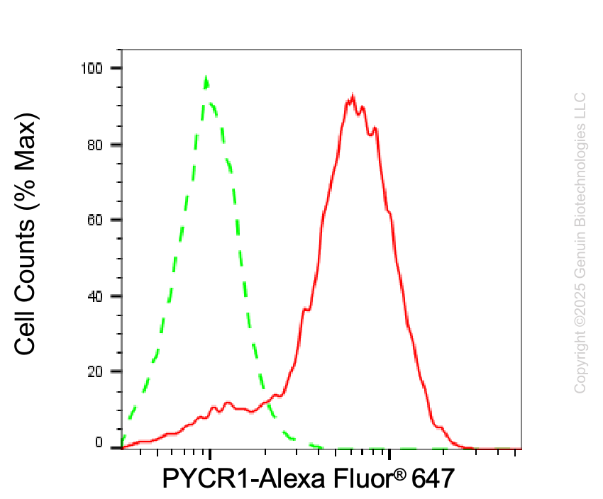
Flow cytometric analysis of PYCR1 expression in HepG2 cells using anti-PYCR1 antibody (Cat#65934, 1:2,000). Green, isotype control; red, PYCR1.
Western blotting analysis using anti-PYCR1 antibody (Cat#65934). PYCR1 expression in wild-type (WT) and PYCR1 shRNA knockdown (KD) HeLa cells with 20 μg of total cell lysates. Hsp90 α serves as a loading control. The blot was incubated with anti-PYCR1 antibody (Cat#65934, 1:2,500) and HRP-conjugated goat anti-mouse secondary antibody (Cat#101, 1:20,000) respectively. Image was developed using NaQ™ ECL Substrate Kit (Cat#716).
Western blotting analysis using anti-PYCR1 antibody (Cat#65934). Total cell lysates (30 μg) from various cell lines were loaded and separated by SDS-PAGE. The blot was incubated with anti-PYCR1 antibody (Cat#65934, 1:2,500) and HRP-conjugated goat anti-mouse secondary antibody (Cat#101, 1:20,000) respectively. Image was developed using NaQ™ ECL Substrate Kit (Cat#716).
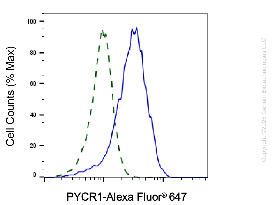
Validation of PYCR1 knockdown using flow cytometry. Wild-type(WT, Blue) and knockdown(KD, Green) HeLa cells were stained with anti-PYCR1 antibody (Cat#65934, 1:2,000) and analyzed using BD flow cytometer.
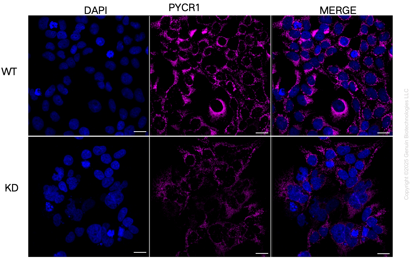
Immunocytochemical staining of HeLa cells using anti-PYCR1 antibody (Cat#65934, 1:1,000), Top panel: wild-type (WT); Bottom panal: PYCR1 shRNA knockdown (KD). Nuclei were stained blue with DAPI; PYCR1 was stained magenta with Alexa Fluor® 647. Scale bar, 20 μm.