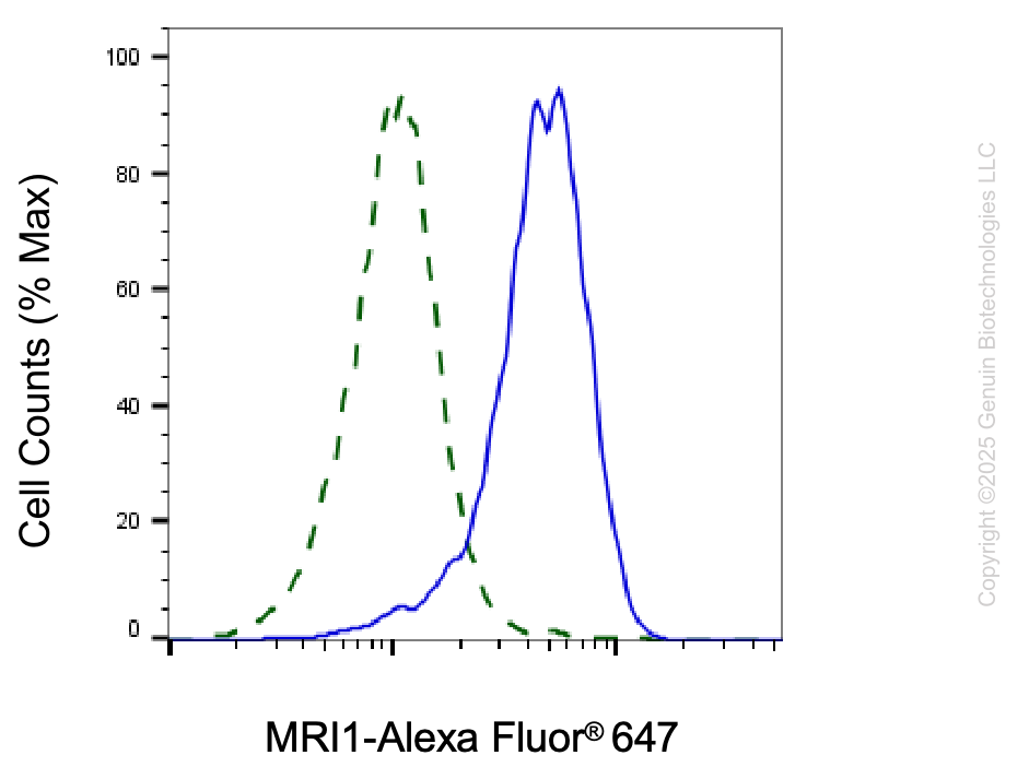
Validation of MRI1 knockdown using flow cytometry. Wild-type(WT, Blue) and knockdown(KD, Green) HepG2 cells were stained with anti-MRI1 antibody (Cat#65829, 1:2,000) and analyzed using BD flow cytometer.
Western blotting analysis using anti-MRI1 antibody (Cat#65829). Total cell lysates (30 μg) from various cell lines were loaded and separated by SDS-PAGE. The blot was incubated with anti-MRI1 antibody (Cat#65829, 1:2,500) and HRP-conjugated goat anti-mouse secondary antibody (Cat#101, 1:20,000) respectively. Image was developed using FeQ™ ECL Substrate Kit (Cat#226).
Western blotting analysis using anti-MRI1 antibody (Cat#65829). MRI1 expression in wild-type (WT) and MRI1 shRNA knockdown (KD) HepG2 cells with 20 μg of total cell lysates. Hsp90 α serves as a loading control. The blot was incubated with anti-MRI1 antibody (Cat#65829, 1:2,500) and HRP-conjugated goat anti-mouse secondary antibody (Cat#101, 1:20,000) respectively. Image was developed using NaQ™ ECL Substrate Kit (Cat#716).
Flow cytometric analysis of MRI1 expression in HAP-1 cells using anti-MRI1 antibody (Cat#65829, 1:2,000). Green, isotype control; red, MRI1.