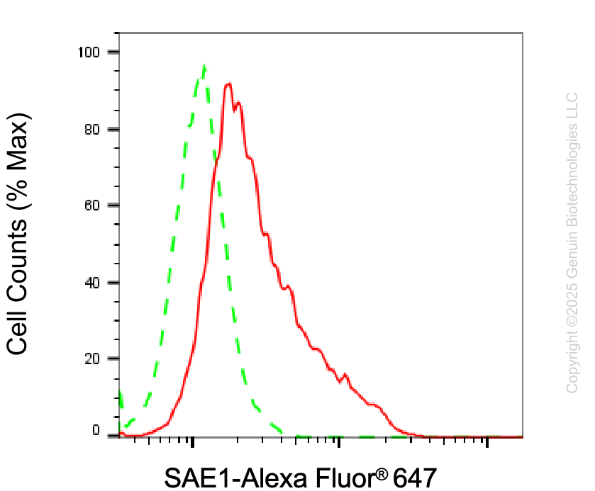
Flow cytometric analysis of SAE1 expression in HAP-1 cells using anti-SAE1 antibody (Cat#5918, 1:2,000). Green, isotype control; red, SAE1.
Western blotting analysis using anti-SAE1 antibody (Cat#5918). Total cell lysates (30 μg) from various cell lines were loaded and separated by SDS-PAGE. The blot was incubated with anti-SAE1 antibody (Cat#5918, 1:2,500) and HRP-conjugated goat anti-mouse secondary antibody (Cat#101, 1:20,000) respectively. Image was developed using FeQ™ ECL Substrate Kit (Cat#226).
Applications Tested: Western blotting (WB), flow cytometry (FCM)
Immunogen
Recombinant protein of human SAE1
Isotype
Mouse IgG1
Storage Buffer
Supplied in PBS (pH 7.4) containing 50% glycerol, and 0.02% sodium azide.
Storage
Store at -20 °C for one year.
Recommended Dilutions
Western Blotting (WB): 1:500-1:2,500 Flow Cytometry (FCM): 1:200-1:2,000
Note
This product is for research use only.
Data
Flow cytometric analysis of SAE1 expression in HAP-1 cells using anti-SAE1 antibody (Cat#5918, 1:2,000). Green, isotype control; red, SAE1.
Western blotting analysis using anti-SAE1 antibody (Cat#5918). Total cell lysates (30 μg) from various cell lines were loaded and separated by SDS-PAGE. The blot was incubated with anti-SAE1 antibody (Cat#5918, 1:2,500) and HRP-conjugated goat anti-mouse secondary antibody (Cat#101, 1:20,000) respectively. Image was developed using FeQ™ ECL Substrate Kit (Cat#226).