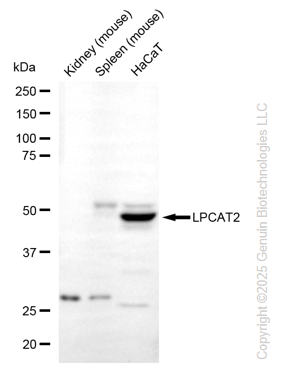
Western blotting analysis using anti-LPCAT2 antibody (Cat#52905). Total lysates (30 μg) were loaded and separated by SDS-PAGE. The blot was incubated with anti-LPCAT2 antibody (Cat#52905, 1:2,500) and HRP-conjugated goat anti-rabbit secondary antibody (Cat#201, 1:20,000) respectively. Image was developed using FeQ™ ECL Substrate Kit (Cat#226).
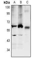
Western blotting analysis using anti-LPCAT2 antibody (Cat#52905). Total lysates were loaded and separated by SDS-PAGE. The blot was incubated with anti-LPCAT2 antibody (Cat#52905, 1:1,000) and secondary antibody respectively. HCT116 (A), DLD (B), mouse colon (C)
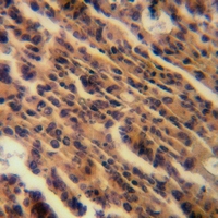
Immunohistochemical analysis of LPCAT2 staining in human colorectal cancer formalin fixed paraffin embedded tissue section. The section was pre-treated using heat mediated antigen retrieval with sodium citrate buffer (pH 6.0). The section was then incubated with the antibody at room temperature and detected using an HRP conjugated compact polymer system. DAB was used as the chromogen. The section was then counterstained with haematoxylin and mounted with DPX.
Applications Tested: Western blotting (WB), immunohistochemistry (IHC)
Immunogen
A synthesized peptide derived from human LPCAT2
Isotype
Rabbit IgG
Storage Buffer
Supplied in PBS (pH 7.3) containing 30% glycerol, and 0.01% sodium azide.
Storage
Store at -20 °C for one year.
Recommended Dilutions
Western Blotting (WB):1:500-1:2,500 Immunohistochemistry (IHC): 1:50-1:100
Note
This product is for research use only.
Data
Western blotting analysis using anti-LPCAT2 antibody (Cat#52905). Total lysates (30 μg) were loaded and separated by SDS-PAGE. The blot was incubated with anti-LPCAT2 antibody (Cat#52905, 1:2,500) and HRP-conjugated goat anti-rabbit secondary antibody (Cat#201, 1:20,000) respectively. Image was developed using FeQ™ ECL Substrate Kit (Cat#226).
Western blotting analysis using anti-LPCAT2 antibody (Cat#52905). Total lysates were loaded and separated by SDS-PAGE. The blot was incubated with anti-LPCAT2 antibody (Cat#52905, 1:1,000) and secondary antibody respectively. HCT116 (A), DLD (B), mouse colon (C)
Immunohistochemical analysis of LPCAT2 staining in human colorectal cancer formalin fixed paraffin embedded tissue section. The section was pre-treated using heat mediated antigen retrieval with sodium citrate buffer (pH 6.0). The section was then incubated with the antibody at room temperature and detected using an HRP conjugated compact polymer system. DAB was used as the chromogen. The section was then counterstained with haematoxylin and mounted with DPX.