• 一抗
  • 二抗
  • 慢病毒
  • 细胞系
  • 裂解物
  • 试剂类

Anti-ZO-1 Rabbit Monoclonal Antibody #52822

一键复制产品信息
Western blotting analysis using anti-ZO-1 antibody (Cat#52822). Total cell lysates (30 μg) from various cell lines were loaded and separated by SDS-PAGE. The blot was incubated with anti-ZO-1 antibody (Cat#52822, 1:2,500) and HRP-conjugated goat anti-rabbit secondary antibody (Cat#201, 1:20,000) respectively. Image was developed using FeQ™ ECL Substrate Kit (Cat#226).
Immunofluorescence-tissue was performed on paraffin-embedded mouse brain sections using an anti-ZO-1 antibody (Cat#52822, 1:200). Antigen retrieval was carried out in sodium citrate buffer (pH 6.0). Nuclei were counterstained with DAPI (blue), and ZO-1 was visualized with Alexa Fluor® 647 (magenta). Protein abundance, based on laser intensity and smart gain settings, was classified as medium. Images were acquired using a Leica Stellaris 5 confocal microscope. Scale bar: 20 µm.
  • 基因名:
    TJP1
  • 货号:
    52822
  • 应用:
    WB, IHC, IF
  • 物种反应性:
    H, M, R
规格:
20 μL,50 μL,100 μL
价格:
¥ 900.00,¥ 1,800.00,¥ 3,300.00
Information
Product Name Anti-ZO-1 Rabbit Monoclonal Antibody
Aliases TJP1; Tight Junction Protein 1; Tight Junction Protein ZO-1; ZO-1; Zona Occludens 1; DKFZp686M05161; MGC133289; Zonula Occludens 1 Protein; Zonula Occludens Protein 1; Zona Occludens Protein 1; ZO1
Background

Gene Name: TJP1

NCBI Gene Entry: 7082

UniProt Entry: Q07157

Application Information

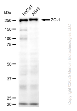
Molecular Weight: Predicted, 195 kDa; observed, 220 kDa

Clonality: Rabbit monoclonal antibody

Clone ID: 25GB7010

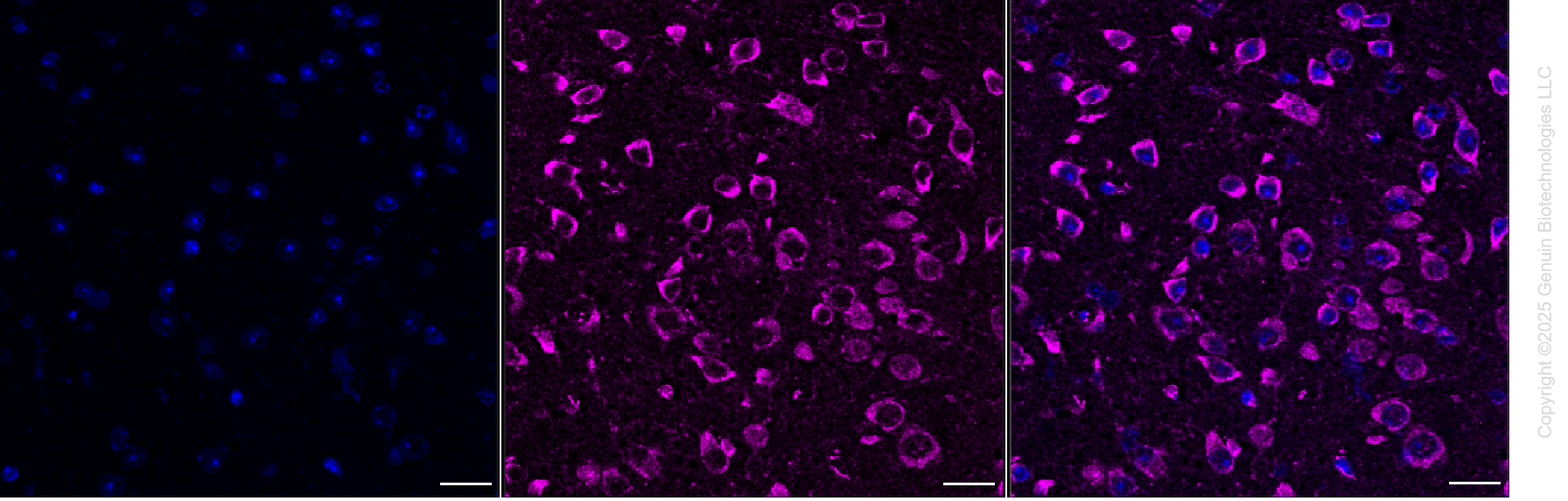
Species Reactivity: Human, mouse, rat

Applications Tested: Western blotting (WB), immunohistochemistry (IHC), immunofluorescence-tissue (IF-Tissue)

Immunogen Recombinant protein of human ZO-1
Isotype Rabbit IgG
Storage Buffer Supplied in 0.01M TBS (pH7.4) containing 50% glycerol, 1% BSA and 0.02% Proclin300.
Storage Store at -20 °C for one year.
Recommended Dilutions Western Blotting (WB): 1:1,000-1:2,500
Immunohistochemistry (IHC): 1:100-1:500
Immunofluorescence-Tissue (IF-Tissue): 1:100-1:200
Note This product is for research use only.
Data
  • Western blotting analysis using anti-ZO-1 antibody (Cat#52822). Total cell lysates (30 μg) from various cell lines were loaded and separated by SDS-PAGE. The blot was incubated with anti-ZO-1 antibody (Cat#52822, 1:2,500) and HRP-conjugated goat anti-rabbit secondary antibody (Cat#201, 1:20,000) respectively. Image was developed using FeQ™ ECL Substrate Kit (Cat#226).
  • Immunofluorescence-tissue was performed on paraffin-embedded mouse brain sections using an anti-ZO-1 antibody (Cat#52822, 1:200). Antigen retrieval was carried out in sodium citrate buffer (pH 6.0). Nuclei were counterstained with DAPI (blue), and ZO-1 was visualized with Alexa Fluor® 647 (magenta). Protein abundance, based on laser intensity and smart gain settings, was classified as medium. Images were acquired using a Leica Stellaris 5 confocal microscope. Scale bar: 20 µm.
Western blotting analysis using anti-ZO-1 antibody (Cat#52822). Total cell lysates (30 μg) from various cell lines were loaded and separated by SDS-PAGE. The blot was incubated with anti-ZO-1 antibody (Cat#52822, 1:2,500) and HRP-conjugated goat anti-rabbit secondary antibody (Cat#201, 1:20,000) respectively. Image was developed using FeQ™ ECL Substrate Kit (Cat#226).
Immunofluorescence-tissue was performed on paraffin-embedded mouse brain sections using an anti-ZO-1 antibody (Cat#52822, 1:200). Antigen retrieval was carried out in sodium citrate buffer (pH 6.0). Nuclei were counterstained with DAPI (blue), and ZO-1 was visualized with Alexa Fluor® 647 (magenta). Protein abundance, based on laser intensity and smart gain settings, was classified as medium. Images were acquired using a Leica Stellaris 5 confocal microscope. Scale bar: 20 µm.
  • 联系电话

    0551-63635518

    0551-65578998

  • 联系邮箱

    support@genuinbiotech.cn

    sales@genuinbiotech.cn

微信公众号

Copyright © 2025 合肥善本生物科技有限公司

All Rights Reserved. 皖ICP备2023000814号