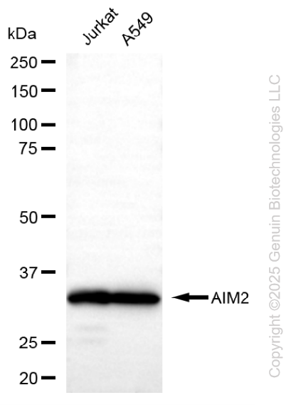
Western blotting analysis using anti-AIM2 antibody (Cat#52539). Total lysates (30 μg) were loaded and separated by SDS-PAGE. The blot was incubated with anti-AIM2 antibody (Cat#52539, 1:2,500) and HRP-conjugated goat anti-rabbit secondary antibody (Cat#201, 1:20,000) respectively. Image was developed using FeQ™ ECL Substrate Kit (Cat#226).
Immunohistochemical analysis of paraffin-embedded human tonsil. Tris-EDTA, pH9.0 was used for antigen retrieval. Antibody was diluted at 1:200 (4℃ overnight). Secondary antibody was diluted at 1:200 (room temperature, 45min).
Immunocytochemical analysis of Hela cell. AIM2 antibody (red) was diluted at 1:200 (4℃ overnight). HSP70 antibody (green) was diluted at 1:200 (4℃ overnight). Goat Anti Rabbit Alexa Fluor 594 was diluted at 1:1,000 (room temperature, 50min). Goat Anti Mouse Alexa Fluor 488 was diluted at 1:1,000 (room temperature, 50min).
Applications Tested: Western blotting (WB), immunohistochemistry (IHC), immunocytochemistry (IC)
Immunogen
A synthesized peptide derived from human AIM2
Isotype
Rabbit IgG
Storage Buffer
Supplied in PBS containing 50% glycerol, and 0.02% sodium azide.
Storage
Store at -20 °C for one year.
Recommended Dilutions
Western Blotting (WB): 1:500-1:2,000 Immunohistochemistry (IHC): 1:100-1:300 Immunocytochemistry (IC): 1:200-1:1,000
Note
This product is for research use only.
Data
Western blotting analysis using anti-AIM2 antibody (Cat#52539). Total lysates (30 μg) were loaded and separated by SDS-PAGE. The blot was incubated with anti-AIM2 antibody (Cat#52539, 1:2,500) and HRP-conjugated goat anti-rabbit secondary antibody (Cat#201, 1:20,000) respectively. Image was developed using FeQ™ ECL Substrate Kit (Cat#226).
Immunohistochemical analysis of paraffin-embedded human tonsil. Tris-EDTA, pH9.0 was used for antigen retrieval. Antibody was diluted at 1:200 (4℃ overnight). Secondary antibody was diluted at 1:200 (room temperature, 45min).
Immunocytochemical analysis of Hela cell. AIM2 antibody (red) was diluted at 1:200 (4℃ overnight). HSP70 antibody (green) was diluted at 1:200 (4℃ overnight). Goat Anti Rabbit Alexa Fluor 594 was diluted at 1:1,000 (room temperature, 50min). Goat Anti Mouse Alexa Fluor 488 was diluted at 1:1,000 (room temperature, 50min).